Endoscopic Anterior Cervical Discectomy & Fusion

Minimally invasive surgery to remove a herniated cervical disc and fuse the spine using an endoscope
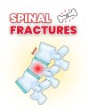
This surgery removes the herniated cervical disc from the front (anterior side) and uses a fusion technique to stabilize the spine.
It treats cervical disc herniation and spinal instability by removing the damaged disc and inserting a cage filled with bone graft to encourage bone fusion and long-term stability.
The procedure uses an endoscopic system to remove the disc and decompress the spinal cord or nerve roots. A cage (usually made of titanium or PEEK) is inserted between the vertebrae and often stabilized with a plate and screws. This allows for rapid recovery, less pain, and greater surgical precision.
Compared to traditional open surgery, this technique uses a much smaller incision (about 2 cm), causes less tissue damage, and has fewer risks and complications.
It's advantages:
- Smaller incision, less pain
- Shorter recovery time
- Lower blood loss
- Minimal infection risk
- Surgical Time
- ️ 2 hours
- Hospital Stay: 4 days / 3 nights
- Incision Size: 2 cm

Brain & Spine Center
Spine Procedures
Others:
Blog


Are you suffering from back pain, neck stiffness, or nerve-related symptoms? Dont ignore the signs. Early diagnosis and expert treatment can prevent long-term complications.